
Contrast-enhanced ultrasound examination is currently the most sensitive imaging procedure with the highest resolution for tissue perfusion examinations. In addition, this method even allows the assessment of blood flow in real time, as was previously only possible with renal function scintigraphy and the use of radioactive markers.
The Ultrasound examination of the kidneys is now a standard method in the hands of nephrologists and Urologiststo give an initial overview of the position, size and shape of the kidneys and, if necessary, a urinary obstruction. Colour-coded Doppler sonography also allows statements to be made about blood flow. For further questions, e.g. the classification of kidney cysts, clarification of a suspected cancer or the diagnosis of a blood vessel occlusion, alternative examinations such as a computer tomogram or magnetic resonance imaging have had to be used up to now. The disadvantage of these methods is the use of contrast media, which in turn are excreted via the kidneys and can themselves damage the kidneys if kidney function is reduced. In addition, these contrast media not only remain in the blood vessels, but also accumulate in the tissues. Iodine-containing X-ray contrast media can also lead to life-threatening thyroid dysfunction.
The contrast medium used for contrast-enhanced ultrasound, on the other hand, consists of a microbubble emulsion. The phospholipid membrane enveloped vesicles the size of a red blood cell contain a medical gas called sulfur hexafluoride, which has virtually no reaction with the body. Therefore, severe hypersensitivity reactions are very rare (risk 1:10,000, in comparison contrast media containing iodine risk 1:100 to 1:1000).
After the intravenous injection of a very small amount of ultrasound contrast medium of less than one millilitre, it floods the renal arteries in 10 to 20 seconds, flows through the segmental and interlobar arteries and then spreads to the approximately half a million renal corpuscles in the renal cortex. This is where the urine filtration takes place. From there, the contrast medium reaches the kidney marrow, which is in great need of oxygen, where the urine is concentrated. After about two minutes, it is hardly possible to detect any more contrast medium bubbles in the kidney. The contrast medium has now been exhaled from the lungs.
Indications for contrast-enhanced ultrasound examination of the kidneys are primarily so-called "all-or-nothing" questions. For example, renal infarctions can be detected very quickly and reliably in patients with cardiac arrhythmias or a tendency to thrombosis. Another important indication is the differentiation of renal cysts into benign, potentially malignant or renal cancer. Initial experience has shown that ultrasound diagnostics are superior to conventional CT diagnostics in the early detection of renal cysts. Therefore, such lesions are discussed together with urologists and radiologists and the further therapeutic procedure (observation or surgical therapy) is determined. In cases of renal pelvic inflammation, focal inflammation and abscesses can be identified. In cases of renal injury or after surgery on the kidney, complications such as vascular fistulas can be reliably identified. A still experimental field is renal function kinetics. In cases of renal function loss, special evaluation software similar to renal function scintigraphy can be used to quantify renal blood flow and show it in a side-by-side comparison.
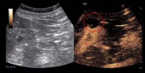
Image: Contrast enhanced sonography. The left picture shows the classic sonography image of the left kidney with a cyst at the upper edge (dark spot). In the right picture the contrast image (brownish) shows the empty cyst with a contrast absorbing structure at the cyst margin at 7 - 9 o'clock. The findings are checked in three months and, if necessary, the cyst must then be removed minimally surgically.


